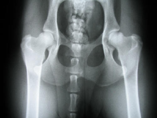
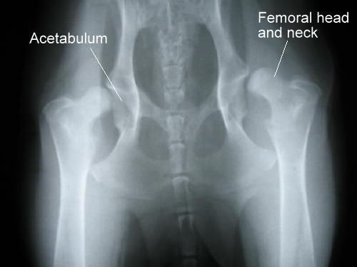
Case 1: Severe bilateral Canine Hip Dysplasia (CHD)
CHD is an abnormality in the development of the hip joint manifested by a shallow acetabulum and a small, misshapen femoral head. Dogs with dysplasia have weak, loose hip joints, weak rear limb muscles and varying degrees of lameness.
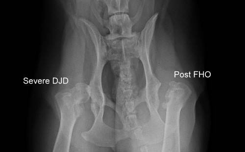
Case 1: Post FHO (dog)
This radiograph is of the same dog as in the previous radiograph and was taken several years after surgical removal of the femoral head and neck from the left femur (Femoral Head and Neck Ostectomy or FHO, right side of image). The right hip (left side of image) shows severe degenerative joint disease or osteoarthritis.
Case 2:
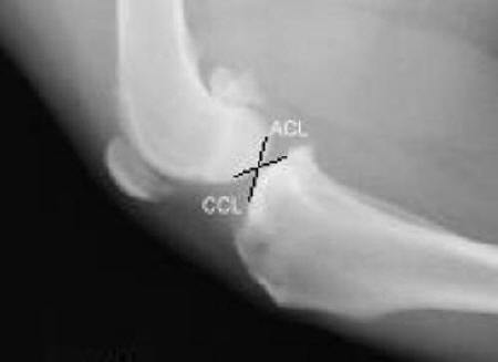
A dog with a torn Anterior Cruciate Ligament (ACL)
Case2: Cruciate Ligaments (dog)
Approximate locations of the Anterior or Cranial Cruciate Ligament (ACL or CrCL) and the Posterior or Caudal Approximate locations of the Anterior or Cranial Cruciate Ligament (ACL or CrCL) and the Posterior or Caudal Cruciate Ligament (PCL or CaCL).
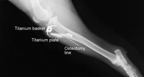
Case 2: Postoperative TTA (dog)
This dog had a Tibial Tuberosity Advancement (TTA) to stabilize its knee with a torn ACL. The procedure involves cutting the tibia (osteotomy line) and then inserting a titanium basket and applying a titanium plate. The gap created in the tibia will fill in with healthy bone.
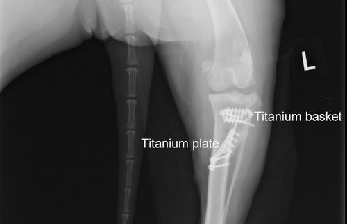
Case 2: Postoperative TTA (dog)
A different view of the same TTA repair as in the previous radiograph.
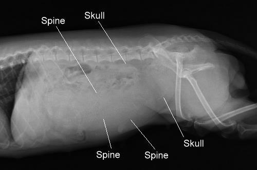
Case 3: Pregnant (dog)
How many puppies?
Case 3: Pregnant (dog)
This dog gave birth to three healthy puppies. The three spines and two of the skulls are indicated. The third skull is not readily visible in this view.
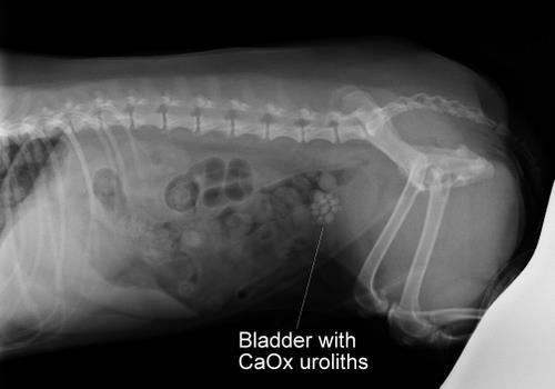
Case 4: Calcium Oxalate uroliths (dog)
A dog with Calcium Oxalate (CaOx) uroliths (bladder stones).
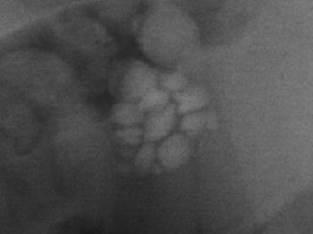
Case 4: Calcium Oxalate uroliths (dog)
This is a magnified view of the previous radiograph. The dog had 15 CaOx uroliths removed from his bladder via cystotomy. This type of urolith is not responsive to dissolution with oral antibiotics and prescription food and must be removed manually.
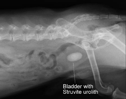
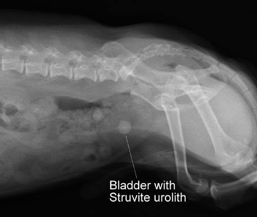
Case 5: Struvite or Magnesium Ammonium Phospate urolith (dog)
This dog presented with dark pink urine. Note the layering of the urolith.
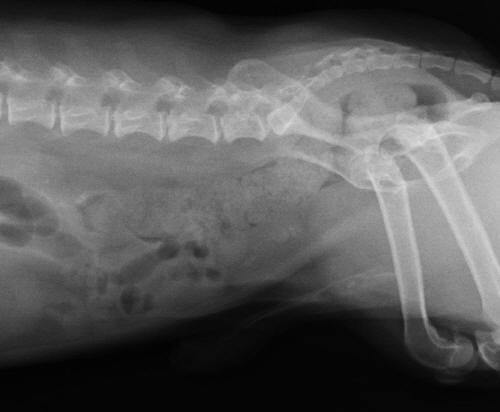
Case 5: Struvite or Magnesium Ammonium Phospate urolith (dog)
This is the same dog as in the previous radiograph. This radiograph was taken one month after the first view. The dog was treated with oral antibiotics and prescription food to encourage dissolution of the urolith. In this image, the urolith is significantly smaller than in the first image.
Case 5: Struvite or Magnesium Ammonium Phospate urolith (dog)
This is the same dog as in the previous two radiographs. This radiograph was taken one month after the previous view. In this radiograph the urolith is almost completely dissolved.
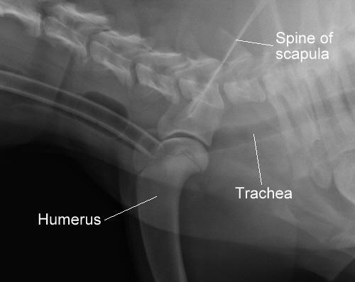
Case 6: Normal shoulder joint (dog)
This dog is anesthetized. The endotracheal tube can be seen as the parallel white lines within the trachea.
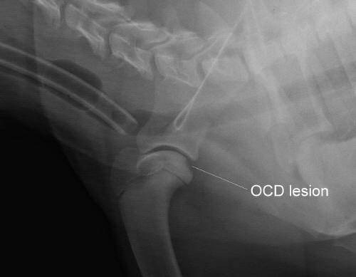
Case 6: Osteochondritis dissecans (OCD) lesion in the shoulder.
This is the same dog as in the previous radiograph. The dog presented with persistent front left limb lameness. In this view an OCD lesion can be seen as an area of cartilage loss on the caudal head of the proximal humerus. OCD is a condition of bone and cartilage inflammation where a piece of cartilage within a joint splits off due to fissure formation in an area of abnormal subarticular cartilage. A flap forms or separates completely and falls into the joint space ("joint mouse"). Treatment involves surgically removing any joint mice and debriding the defect in the cartilage to stimulate normal cartilage growth.
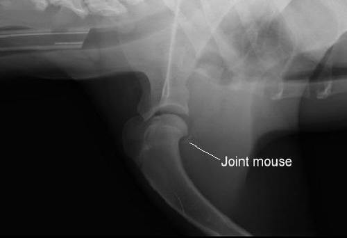
Case 6: Osteochondritis dissecans (OCD) lesion in the shoulder.
A different dog than in the previous two radiographs with shoulder OCD and a calcified flap or "joint mouse" in the caudal cul de sac.
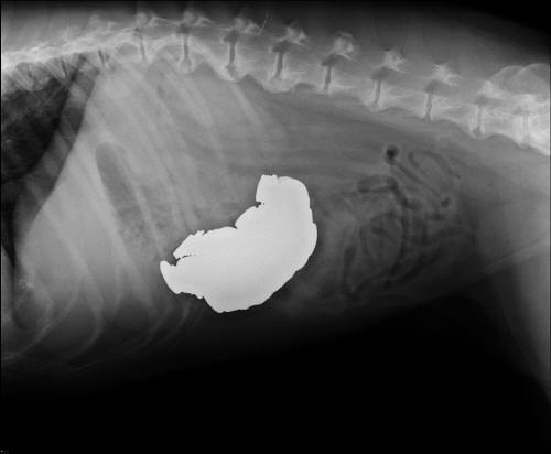
Case 7: Gastric foreign bodies (dog)
This dog had $13.19 worth of loose coins surgically removed from her stomach. If you look closely you can see the edges of individual coins. Note that the coins (metal) show up as bright white on the radiograph because they are so radiodense that x-rays are unable to penetrate them to reach the film.
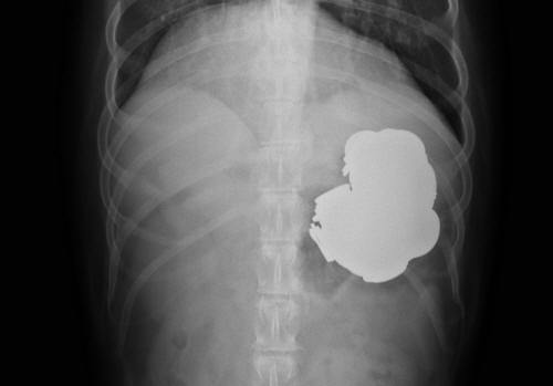
Case 7: Gastric foreign bodies (dog)
A different view of the same dog as in the previous radiograph.
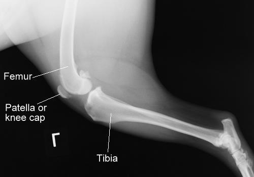
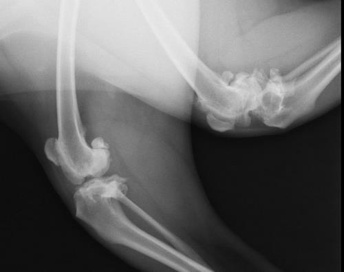
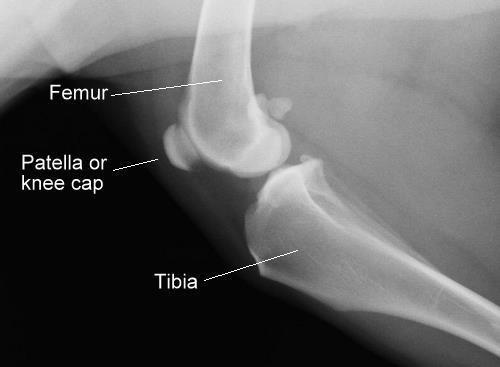
Case 8: Normal knee (dog)
Note the smooth surfaces of the bones.
Case 8: Severe Degenerative Joint Disease or Osteoarthritis.
Note the dramatic degenerative changes to both knees in this dog compared to the previous radiograph.
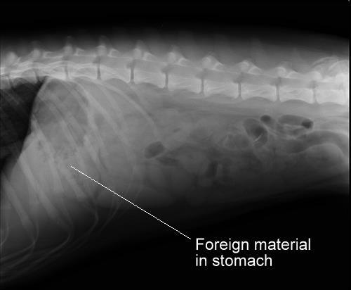
Case 9: Gastric foreign material (dog)
Foreign material is present in this dog's stomach outflow region (pylorus). The dog presented for multiple episodes of vomiting over the previous two days. A chewed up sock was removed during surgical exploration of her stomach and small intestine.
Case 10: Normal knee (dog)
Note the smooth surfaces of the bones as well as the uniform texture of the cortices (outer layers of the bones) and medullary cavities (inner portions of the bones).
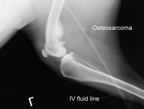
Case 10: Osteosarcoma (dog)
This is the same dog as in the previous radiograph. It presented for persistent lameness on the left rear leg. An aggressive, malignant bone tumor, osteosarcoma, can be seen on the caudal aspect of the distal femur. Osteosarcoma causes bone loss (osteolysis, irregular texture within the medullary cavity) as well as proliferation of abnormal cortical bone at the site of the tumor. The most common sites for osteosarcoma to occur are the proximal humerus and the distal radius (away from the elbow) and the distal femur and the proximal tibia (towards the knee). Large and giant breed dogs are more commonly affected than small to medium breed dogs. Cats can be affected as well.
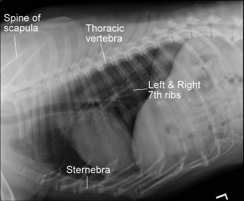
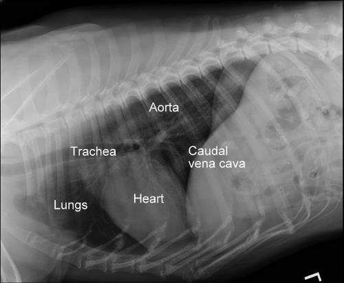
Case 11: Normal thorax (older dog)
Boney structures are labeled in this view.
Case 11: Normal thorax (older dog)
Soft tissue structures are labeled in this view. The air-filled trachea and lungs appear dark or black on radiographs. The multiple white lines within the lung fields are arteries, veins and airways.
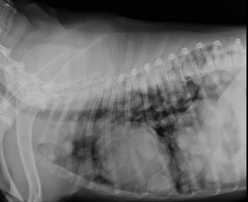
Case 11: Pulmonary neoplasia (dog)
This dog's lungs are diffusely affected by countless, nodular tumors. The primary rule out is primary or secondary neoplasia or cancer of the lungs although a fungal infection is possible
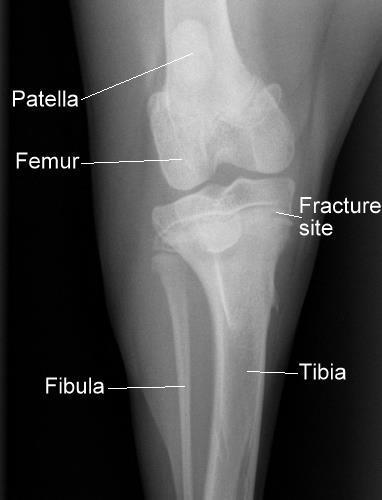
Case 12: Normal knee (puppy)
Note the growth plates for the tibial tuberosity on the left and the tibial condyles on the right. These growth plates will fill in as the puppy matures.
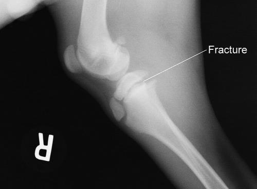
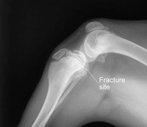
Case 12: Salter-Harris Type I fracture (puppy)
This is the same puppy as in the previous radiograph. In this view, a Salter-Harris Type I fracture of the right proximal tibial condylar growth plate can be seen as a wide gap in the growth plate compared to the normal knee.
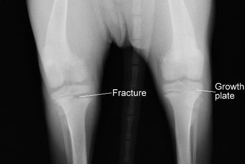
Case 12: Salter-Harris Type I fracture (puppy)
This is a different view of the same puppy as in the previous two views. This view allows direct comparison between the fractured growth plate (left side of the image) and the normal growth plate (right side of the image).
Case 12: Salter-Harris Type I fracture (puppy)
Another view showing the healed fracture site after four weeks of splinting.
Case 12: Salter-Harris Type I fracture (puppy)
This view of the same puppy is after approximately four weeks of splinting of the affected leg to immobilize the fracture site. The fracture has healed appropriately. However, there is some mild bowing of the tibia (proximal tibial valgus) due to the injury to the growth plate. This condition is not expected to cause serious issues for the puppy as it matures
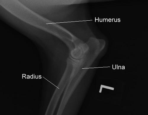
Case 13: Ulnar ostectomy pre-operative (dog)
This dog presented for persistent front left leg lameness. She was diagnosed with a possible OCD lesion (see Case 6) of the left elbow, a likely fractured medial coronoid process (FCP) of the proximal ulna and likely subluxation (partial dislocation) of the elbow due to a growth disparity between the radius and ulna.
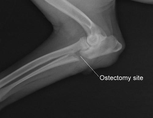
Case 13: Ulnar ostectomy post-operative (dog)
This is the same limb of the same dog as in the previous radiograph following proximal ulnar ostectomy in which a segment of bone was surgically removed to compensate for a growth disparity between the radius and ulna. In addition to the ulnar ostectomy, a large OCD lesion was removed from the distal medial humerus. No FCP lesion was identified.
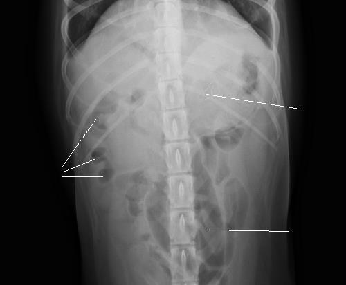
Case 14: Gastrointestinal obstruction (dog)
This dog presented for several days of vomiting, lethargy and trembling. She was known to have recently eaten a dish towel. This radiograph shows uniform fine material suggestive of fabric and a thin twist tie wire in the stomach and marked distention of the small intestine with gas. The black line across the radiograph is an artifact.
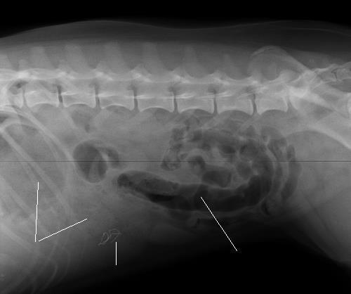
Case 14: Gastrointestinal obstruction (dog)
This is a different view of the same dog as in the previous radiograph. On the left side of the radiograph there are several "tear drop-shaped" gas pockets in the small intestine strongly suggestive of a linear foreign body. During surgery, an incision into her stomach (gastrotomy) and two incisions into her small intestines (enterotomies) were required to remove several clumps of dish towel fabric with string connections between and an orange twist tie.
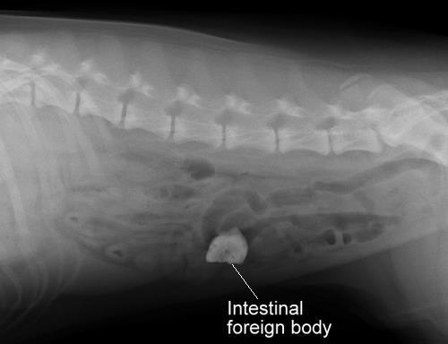
Case 15: Intestinal foreign body (dog)
This dog presented for 2 days of decreased appetite and inability to hold down food or water. He had a history of eating wood chips and may have eaten a rock. A firm object was palpable in his abdomen. The lateral radiograph revealed a radiodense object in his intestines with a mottled appearance suggested a piece of rawhide. The owners declined surgery to remove the foreign body. After a couple of days of intestinal lubricant the dog passed a rock wrapped in grass.